Genome-wide association study identifies two
susceptibility loci for osteosarcoma
Osteosarcoma is the most common primary bone malignancy of adolescents and young adults. To better understand the genetic etiology of osteosarcoma, we performed a multistage genome-wide association study consisting of 941 individuals with osteosarcoma (cases) and 3,291 cancer-free adult controls of European ancestry. Two loci achieved genome-wide significance: a locus in the GRM4 gene at 6p21.3 (encoding glutamate receptor metabotropic 4; rs1906953; P = 8.1 × 10⁻⁹) and a locus in the gene desert at 2p25.2 (rs7591996 and rs10208273; P = 1.0 × 10⁻⁸ and 2.9 × 10⁻⁷, respectively). These two loci warrant further exploration to uncover the biological mechanisms underlying susceptibility to osteosarcoma.
LINK: https://www.nature.com/articles/ng.2645
Vários fatores influenciam na demora para que o paciente com diagnóstico de osteossarcoma seja encaminhado para tratamento a um serviço especializado. Objetivo:conhecer o itinerário terapêutico de pacientes com osteossarcoma desde os primeiros sinais e sintomas até serem atendidos em um serviço de saúde especializado. o referencial teórico utilizado foi o modelo de cuidado à saúde proposto por Kleinman, que compreende os subsistemas: profissional, familiar e popular. Método: trata-se de um estudo descritivo, de abordagem mista, realizado no instituto de oncologia Pediátrica (ioP), são Paulo. Resultados: Participaram do estudo 16 pacientes, sendo sete do sexo feminino (43,75%) e nove do masculino (56,25%), com idade entre 12 e 23 anos. os sinais e sintomas: dor, edema e dificuldade de locomoção, apresentados por 43,75% dos pacientes, foram os principais motivadores da procura por atendimento de saúde. o sistema familiar foi o primeiro acionado na busca de solução do problema. Posteriormente, metade dos pacientes (oito), juntamente com a família, procurou um serviço de saúde e os outros 50% realizaram autotratamento. o tempo médio desde o aparecimento dos sinais e sintomas iniciais até a chegada a um serviço especializado foi de seis meses. Conclusão: Foi observada uma forte influência do subsistema familiar na tomada de decisão sobre a escolha do itinerário terapêutico dos pacientes deste estudo. entretanto, falhas nos diferentes níveis de atenção à saúde contribuíram para o atraso significativo no estabelecimento do diagnóstico.
Objetivo: Avaliar os resultados obtidos com o uso de protocolo fisioterapêutico especialmente
desenvolvido para pacientes submetidos à cirurgia de ressecção tumoral ao redor do joelho e
reconstrução com endoprótese não convencional. Métodos: Foram analisados,
prospectivamente, 23 pacientes submetidos à ressecção do tumor ósseo ao redor do joelho e
substituição por endoprótese total não convencional. A média de idade dos pacientes foi de 18
anos. Todos os pacientes apresentavam osteossarcoma no resultado anatomopatológico, sendo
13 (56,5%) com localização na extremidade distal do fêmur (grupo I) e 10 (43,5%) na proximal de
tíbia (grupo II). O tratamento fisioterapêutico iniciouse logo após a definição do diagnóstico,
com duração mínima de seis meses de pósoperatório. Resultados: No grupo I o arco de movimento apresentou média de 112°; no grupo II, de 87°. O grau de força muscular do
quadríceps variou de 4 a 5 no grupo I e de 2 a 5 no grupo II. Todos os demais músculos
apresentaram grau 5 de força muscular. A análise observacional da marcha foi normal em 100%
dos pacientes do grupo I e 4 dos 10 pacientes do grupo II apresentaram claudicação. Conclusão: Os pacientes com tumor ósseo em extremidade distal do fêmur apresentaram resultado melhor
do que os acometidos em extremida.
Results of the Brazilian Osteosarcoma Treatment
Group Studies III and IV: Prognostic Factors and
Impact on Survival.
From the Instituto de Oncologia Pedia´ trica, Grupo de Apoio ao Adolescente e a criança com Câncer/UniversidadeFederal de São Paulo; Pediatrics Department, Hospital do Câncer; Hospital das Clínicas de São Paulo-Faculdade de Medicina da Universidade de SãoPaulo; Santa Casa de Misericórdia de São Paulo; Departments of Orthopedics and Pathology, Universidade Federal de
São Paulo-Escola Paulista de Medicina, São Paulo; Hospital de Clínicas de Porto Alegre, Porto Alegre, Brazil; and the Children’s Hospital of Los Angeles, Los Angeles, CA.
Submitted July 22, 2005; accepted December 16, 2005.
Authors’ disclosures of potential conflicts of interest and author contributions are found at the end of this article.
Address reprint requests to Antonio Sérgio Petrilli MD, PhD, Instituto de Oncologia Pediátrica, Grupo de Apoio ao Adolescente e a Criança com Câncer/Universidade Federal de São Paulo, Rua Botucatu 743, Vila Clementino, 04023-062 São Paulo, Brazil; e-mail: iopepm@dialdata.com.br.
© 2006 by American Society of Clinical Oncology
Activity of Intraarterial Carboplatin as a Single Agent in the Treatment of
Newly Diagnosed Extremity Osteosarcoma

Ototoxicity evaluation in medulloblastoma patients treated with involved field boost using intensity-modulated radiation therapy (IMRT): a retrospective review
Purpose
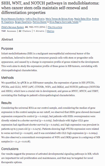
Infant medulloblastoma (MB) is a malignant neuroepithelial embryonal tumor of the cerebellum, believed to derive from precursor granule cells with stem or progenitor cells appearance, and caused by a change in expression profile of genes related to the development. This work aims to study the expression profile of these genes in MB tumors, correlating with clinicopathological characteristics.
Methods
We quantified, by qPCR in 40 MB tumor samples, the expression of genes in HH (PTCH1, PTCH2, and GLI1), WNT (APC, CTNNB1, WIF1, and DKK2), and NOTCH pathways (NOTCH2 and HES1), which have a crucial role in development, and genes as MYCC, MYCN, and TERT, correlating this findings to patient’s clinicopathological characteristics.
Results
Considering the universal RNA as our control sample, and considering the median of gene expression in the control samples as our cutoff, we observed that HES1 gene showed decreased expression compared to control (p = 0.0059), but patients with HES1 overexpression were directly related to a shorter survival (p = 0.0165). Individuals with higher GLI1 gene expression had significant shorter survival (p = 0.0469), and high expression was prevalent in patients up to 5 years old (p = 0.0479). Patients showing high PTCH2 expression were related to worse survival (p = 0.0426), and it was correlated with GLI1 high expression (p = 0.0094). We also observed a concomitant overexpression of WIF1 and DKK2 genes in a subgroup of MB samples (n = 11, p = 0.0118).
Conclusions
Our results suggest the presence of activated developmental signaling pathways in MB, which are important for cell proliferation and maintenance, and that may be targeted for novel therapeutic options.
LINK: https://link.springer.com/article/10.1007%2Fs00381-014-2403-x

Medulloblastoma is the most common malignant tumors of central nervous system in the childhood. The treatment is severe, harmful and, thus, has a dismal prognosis. As PRAME is present in various cancers, including meduloblastoma, and has limited expression in normal tissues, this antigen can be an ideal vaccine target for tumor immunotherapy. In order to find a potential molecular target, we investigated PRAME expression in medulloblastoma fragments and we compare the results with the clinical features of each patient. Analysis of gene expression was performed by real-time quantitative PCR from 37 tumor samples. The Mann-Whitney test was used to analysis the relationship between gene expression and clinical characteristics. Kaplan-Meier curves were used to evaluate survival. PRAME was overexpressed in 84% samples. But no statistical association was found between clinical features and PRAME overexpression. Despite that PRAME gene could be a strong candidate for immunotherapy since it is highly expressed in medulloblastomas.
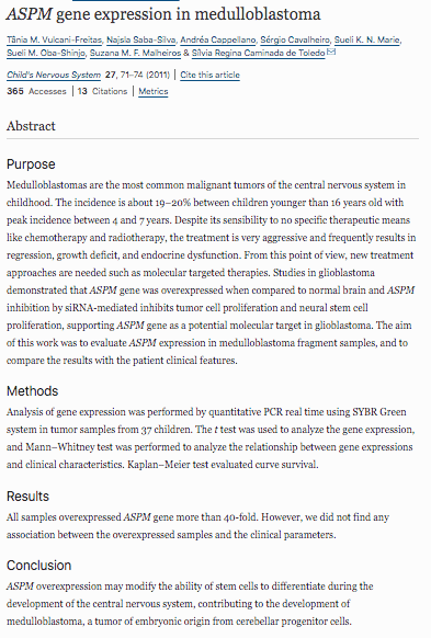
Medulloblastomas are the most common malignant tumors of the central nervous system in childhood. The incidence is about 19–20% between children younger than 16 years old with peak incidence between 4 and 7 years. Despite its sensibility to no specific therapeutic means like chemotherapy and radiotherapy, the treatment is very aggressive and frequently results in regression, growth deficit, and endocrine dysfunction. From this point of view, new treatment approaches are needed such as molecular targeted therapies. Studies in glioblastoma demonstrated that ASPM gene was overexpressed when compared to normal brain and ASPM inhibition by siRNA-mediated inhibits tumor cell proliferation and neural stem cell proliferation, supporting ASPM gene as a potential molecular target in glioblastoma. The aim of this work was to evaluate ASPM expression in medulloblastoma fragment samples, and to compare the results with the patient clinical features.
LINK: https://link.springer.com/article/10.1007%2Fs00381-010-1252-5
Aberrant expression of stem cell-related genes in tumors may confer more primitive and aggressive traits affecting clinical outcome. Here, we investigated expression and prognostic value of the neural stem cell marker CD133, as well as of the pluripotency genes LIN28 and OCT4 in 37 samples of pediatric medulloblastoma, the most common and challenging type of embryonal tumor. While most medulloblastoma samples expressed CD133 and LIN28, OCT4 expression was found to be more sporadic, with detectable levels occurring in 48% of tumors. Expression levels of OCT4, but not CD133 or LIN28, were significantly correlated with shorter survival (P ≤ 0.0001). Median survival time of patients with tumors hyperexpressing OCT4 and tumors displaying low/undetectable OCT4 expression were 6 and 153 months, respectively. More importantly, when patients were clinically stratified according to their risk of tumor recurrence, positive OCT4 expression in primary tumor specimens could discriminate patients classified as average risk but which further deceased within 5 years of diagnosis (median survival time of 28 months), a poor clinical outcome typical of high risk patients. Our findings reveal a previously unknown prognostic value for OCT4 expression status in medulloblastoma, which might be used as a further indicator of poor survival and aid postoperative treatment selection, with a particular potential benefit for clinically average risk patients.
LINK: https://link.springer.com/article/10.1007%2Fs11060-011-0647-9
Purpose Gliomas represent the most frequent central nervous system (CNS) tumors in children and adolescents. However,
therapeutic strategies for these patients, based on tumor molecular profile, are still limited compared to the wide range of
treatment options for the adult population. We investigated molecular alterations, with a potential prognostic marker and
therapeutic target in gliomas of childhood and adolescence using the next-generation sequencing (NGS) strategy.
Methods We selected 95 samples with initial diagnosis of glioma from patients treated at Pediatric Oncology Institute-GRA
ACC/UNIFESP. All samples were categorized according to the 2021 World Health Organization Classification of Tumors
of the CNS, which included 39 low-grade gliomas (LGGs) and 56 high-grade gliomas (HGGs). Four HGG samples were
classified as congenital glioblastoma (cGBM). NGS was performed to identify somatic genetic variants in tumor samples
using the Oncomine Childhood Cancer Research Assay® (OCCRA®) panel, from Thermo Fisher Scientific®.
Results Genetic variants were identified in 76 of 95 (80%) tumors. In HGGs, the most common molecular alteration detected
was H3F3A c.83A>T variant (H3.3 K27M) and co‐occurring mutations in ATRX, TP53, PDGFRA, MET, and MYC genes
were also frequently observed. One HGG sample was reclassified as supratentorial ependymoma ZFTA-fusion positive after
NGS was performed. In LGGs, four KIAA1549–BRAF fusion transcripts were detected and this alteration was the most recurrent genetic event and favorable prognostic factor identified. Additionally, genetic variants in ALK and NTRK genes, which
provide potential targets for therapy with Food and Drug Administration-approved drugs, were identified in two different
cases of cGBM that were classified as infant-type hemispheric glioma, a newly recognized subgroup of pediatric HGG.
Conclusion Molecular profiling by the OCCRA® panel comprehensively addressed the most relevant genetic variants in
gliomas of childhood and adolescence, as these tumors have specific patterns of molecular alterations, outcomes, and effectiveness to therapies
Single agent vinorelbine in pediatric patients with progressive optic pathway glioma
The management of progressive unresectable low-grade glioma remains controversial. Treatment options have included radiotherapy, and more recently chemotherapy, usually following an initial period of observation. Within this context, we evaluated vinorelbine, a semi-synthetic vinca alkaloid that has shown evidence of activity against glioma. From July 2007 an institutional protocol with vinorelbine (30 mg/m2 days 0, 8, 22) for a total of 18 cycles, has been conducted at IOP/GRAACC/UNIFESP for children with optic pathway glioma (OPG). The main objectives were clinical and radiological response, as well as toxicity profile. Twenty-three patients with progressive OPG with a mean age of 69 months (4–179) were enrolled. Three patients had a diagnosis of neurofibromatosis type 1. Twenty-two patients were assessable for response with an overall objective response rate of 63 %, with eight patients showing stable disease. The most important toxicity was hematologic (grade III/IV neutropenia) observed in four patients. Gastrointestinal toxicity (grade I/II vomiting) was observed in seven patients and only 1 patient showed grade I peripheral neuropathy. The median progression-free survival (PFS) was 33 months (6.9–69) with a 3 and 5 year PFS of 64 ± 19 and 37 ± 20 %, respectively, for an overall 3 and 5 year-survival of 95 ± 10 %. This study suggests that vinorelbine may be an interesting option for pediatric low-grade gliomas, showing low toxicity profile and providing a good quality of life for patients with such chronic disease.
LINK: https://link.springer.com/article/10.1007/s11060-014-1652-6
Diffuse Intrinsic Brainstem Tumor in an Infant
Brainstem gliomas constitute 10% to 20% of all pediatric tumors of the central nervous system, and diffusely infiltrative brainstem gliomas are the most common brainstem tumors associated with a poor prognosis. A small subset of these tumors is benign, showing low-grade features on histology. The role of chemotherapy in the management of these tumors is ill defined, especially in the neonates. There are anecdotal reports of spontaneous remission, but the natural history of these tumors does not support a wait-and-see approach. Thus, we report a successful experience of chemotherapy in a 4-month-old girl with a diffuse brainstem fibrillary astrocytoma, treated with vinorelbine (30 mg/m2/d on days 0, 8, and 22), a vinca alkaloid that has shown activity against glioma. Our experience suggests that vinorelbine may be effective in pediatric low-grade gliomas as this patient showed significant clinical improvement over a short period of time.
LINK: https://journals.lww.com/jpho-online/Abstract/2011/03000/Diffuse_Intrinsic_Brainstem_Tumor_in_an_Infant___A.9.aspx
Objective: To outline the epidemiological profile and prognosis for Ewing’s sarcoma in the Brazilian population. Material and Methods: The medical records of 64 patients with intraosseous Ewing’s sarcoma who were treated at the Pediatric Oncology Institute, IOP-GRAACC-Unifesp, between 1995 and 2010, were retrospectively evaluated. Results: The statistical analysis on the data obtained did not correlate factors such as sex, trauma, pathological fracture and time taken for case diagnosis with the treatment outcome. Factors such as initial metastasis, lung metastasis, tumor site, age, recurrence and type of surgery showed results corroborating what has been established in the literature. Conclusion: The prognosis in cases of Ewing’s sarcoma was mainly influenced by the presence of metastases at the time of diagnosis.
Background
Large cooperative group studies have shown the efficacy of risk-adapted treatment for Ewing sarcoma. However, validation and local adaptation by National cooperative groups is needed. A multicenter protocol to determine the efficacy and safety of a risk-adapted intensive regimen was developed by the Brazilian cooperative group.
Procedure
Patients <30 years old with Ewing sarcoma were eligible. Induction chemotherapy consisted of two cycles of ICE (ifosfamide, carboplatin, and etoposide) followed by two cycles of VDC (vincristine, doxorubicin, and cyclophosphamide), followed by local control. Patients with low risk (LR) disease (localized resectable with normal LDH) received 10 additional alternating courses of IE with VDC. For patients with high-risk (HR) disease (unresectable, pelvic, metastatic, or high LDH), two additional cycles of ICE were given.
Results
One-hundred seventy five patients (39% metastatic) were enrolled. Fifty-two patients (29.7%) were LR and 123 (70.3%) were HR. Overall response rate at end of induction was 27.4%. Five-year event-free survival (EFS) and overall survival (OS) estimates were 51.4% and 54.4%, respectively. Patients with localized disease had better outcomes than patients with metastases (5-year EFS 67.9% vs. 25.5%, and 5-year OS 70.3% vs. 29.1%, respectively). On multivariate analysis, the presence of metastatic disease was the only prognostic factor (P < 0.01).
Conclusion
The VDC/ICE protocol was feasible, and considering the high tumor burden in our population, resulted in comparable results to those reported by cooperative groups in high-income countries. Further adaptation to maximize efficacy and minimize toxicity will be required. Pediatr Blood Cancer 2015;62:1747–1753. © 2015 Wiley Periodicals, Inc.
LINK: https://onlinelibrary.wiley.com/doi/abs/10.1002/pbc.25562
Desmoplastic infantile ganglioglioma (DIG; also known as desmoplastic infantile astrocytoma, DIA) is a rare, World Health Organization (WHO) grade-I central nervous system tumor that most often arises in very young children, often with rapidly enlarging head circumference. We reviewed the records of all infants diagnosed with pathology-confirmed DIG/DIA at Mayo Clinic from 1997 to 2016. Immunohistochemical staining was performed on tumor specimens to assess expression of IDH1 R132H, BRAF V600E, H3K27me3 and INI1. Seven patients were included in this study: age range was 2 - 9 months, and four were male. Six patients presented with rapidly enlarging head circumference, and one presented with isolated seizures. On immunohistochemical analysis, all tumors were negative for R132H-mutant IDH1. INI1 expression was retained in all specimens, and BRAF V600E expression was negative or only very focally positive. H3K27me3 expression was partly lost in four tumors. Three tumors were located in the suprasellar region, and four were located in the lateral cerebral hemispheres. All patients underwent surgery for tumor resection; a gross total resection was achieved in the lateral cerebral tumors, and a subtotal resection was achieved in the suprasellar tumors. At 2-year follow-up, none of the patients with a hemispheric tumor developed long-term sequelae from their surgery, but each patient with a supresellar tumor had a neurologic or endocrinologic deficit from tumor progression or surgery, including visual impairment, panhypopituitarism and/or hemiparesis. These results highlight the importance of tumor location in prognosticating these challenging tumors, suggesting that suprasellar location is associated with a poorer prognosis.
Low-grade astrocytomas comprise about 30 % of the central nervous system tumors in children. Several investigations have searched a correlation between the BRAF gene fusions alterations and mutations at IDH1 and IDH2 genes in low grade pediatric astrocytomas. This study identified the expression of KIAA1549–BRAF fusion gene and BRAF V600E mutation, mutations at exon 4 of the IDH1 and IDH2 genes in samples of pilocytic astrocytomas (PA) and grade-II astrocytomas (A-II) pediatric patients. The correlation between these alterations and the clinical profile of the patients was also evaluated. Eighty-two samples of low-grade astrocytomas (65 PA and 17 A-II) were analyzed by PCR and sequencing for each of the targets identified. We identified the KIAA1549–BRAF fusion transcript in 45 % of the samples. BRAF V600E and BRAFins598T mutations were detected in 7 and 1 % of the samples, respectively. Mutations in the R132/R172 residues of the IDH1/IDH2 genes were detected in only two samples, and the G105G polymorphism (rs11554137:C>T) was identified in ten patients. Additionally, we observed two mutations out of the usual hotspots at IDH1 and IDH2 genes. We observed a smaller frequency of mutations in IDHs genes than previously described, but since the prior studies were composed of adult or mixed (adults and children) samples, we believe that our results represent a relevant contribution to the growing knowledge in low grade childhood astrocytomas.
LINK: https://link.springer.com/article/10.1007%2Fs11060-014-1398-1
INTRODUCTION
Management of brain tumors in neonates and infants is very challenging.
OBJECTIVE
To analyze the several treatment strategies and outcomes of brain tumors in the first year of life in a single institution.
METHODS
The authors retrospectively evaluated 63 infants with brain tumors treated between 2007 and 2017, at IOP/GRAACC/UNIFESP. Data regarding initial clinical presentation, treatment modalities and outcomes were collected.
RESULTS
From 63 patients treated, 61 were eligible for evaluation and 2 were excluded for loss of follow-up. Thirty were girls and 31 boys. The mean age at treatment was 6 months (range: 1 day-12 months). Twenty-nine babies presented with signs and symptoms of intracranial hypertension, 11 babies with epileptic seizures and 8, cranial nerves deficits. Forty-five tumors were located in the supratentorial compartment and 16 were infratentorially. Nine patients were diagnosed with tuberous sclerosis, 2 with neurofibromatosis type 1, 2 diagnosed with Li Fraumeni and 1 with Gorlin syndrome. The most common histological types were: 11 rhabdoid teratoid, 9 low grade astrocytoma, 5 choroid plexus carcinoma and 5 glioblastoma. Surgery is the treatment of choice. Ten patients underwent more than one surgery (2 to 6 resections) Eight deaths occurred. The mean follow-up was 3y10m (range: 2 days- 7y9m).
CONCLUSIONS
Gross total resection is the goal of surgical treatment, but sometimes it is impossible in the first approach. To decrease the high intraoperative mortality of malignant tumors, these patients can undergo as many surgeries as necessary for total tumor resection, alternating with cycles of chemotherapy.
Craniopharyngioma is a sellar/suprasellar benign tumor whose
aggressiveness may imply in endocrine disturbances (hypothalamic obesity and hormone deficiencies). Fifty-seven patients were evaluated according to clinical characteristics, hypothalamic involvement, type of treatment, anthropometric variables, adiposity indexes (body mass index Z score category at diagnosis and post-treatment, total body fat, visceral adipose
tissue, and metabolic syndrome components) and analyzed through multiple regression and logistic models. Patients were stratified according to growth hormone deficiency and recombinant human growth hormone use. Mean ages at diagnosis and at study evaluation were 9.6 and 16.6 years old, respectively. A set of 43/57 (75.4%) patients presented with important hypothalamic involvement, 24/57 (42.1%) received surgical treatment and cranial radiotherapy, and 8/57 (14 %)
interferon-α exclusively. Fifty-five patients (96.5%) were considered growth hormone deficient, and 26/57 (45.6%) grew despite no recombinant human growth hormone replacement therapy. At diagnosis, 12/57 (21%) patients were obese, and 33/57 (57.9%) at study evaluation, and after 3.2 years (median) post first therapy. There was no influence of height Z score on body mass index Z score. Body mass index Z score at diagnosis positively influenced body mass index Z score, total body fat, waist circumference and the presence of the metabolic syndrome post-treatment. Replacement of recombinant human growth hormone decreased total body fat and visceral adipose tissue. Craniopharyngioma patients worsened body mass index Z score category 3.2 years (median) after first treatment. Body mass index Z score increased due to real weight gain, without height decrease. Replacement of recombinant human growth hormone had beneficial effect on adiposity.
Objectives
The aim of this study was to verify whether intracystic injections of alpha-Interferon (IFN-α) in cystic craniopharyngiomas were able to reduce the tumor by activating the Fas apoptotic pathway.
Materials and methods
Twenty-one patients with cystic craniopharyngiomas treated at the Pediatric Oncology Institute (IOP/GRAACC) of Federal University of São Paulo were submitted to intracystic chemotherapy with IFN-α. The tumor sizes of all patients were monitored and the apoptotic factor soluble FasL (sFasL) concentration was determined by ELISA (enzyme-linked immunosorbent assay) in tumor fluid samples from eight patients.
Results
There was a complete reduction in 11 patients, a partial response in seven, and a minor response in three patients. The concentration of sFasL was increased in all the eight patients examined concomitantly with the tumor size reduction.
Conclusions
Our data demonstrated that the IFN-α was able to induce Fas-mediated apoptosis together with a reduction in the tumor size; such an observation may suggest the importance to investigate still unexplored mechanisms to be exploited in craniopharyngioma therapy.
LINK: https://link.springer.com/article/10.1007/s00381-007-0409-3

To assess whether the cystic craniopharyngiomas can be controlled with the
use of intratumoral applications of interferon alpha. Method: Nineteen patients with the
diagnosis of cystic craniopharyngioma were treated with intratumoral chemotherapy with
interferon alpha from January 2002 to April 2006. All patients underwent placement of
an intracystic catheter connected to an Ommaya reservoir. Through this reservoir were
made applications during chemotherapy cycles. Each cycle corresponded to application
of 3,000,000 units of interferon alpha three times per week on alternate days totalizing
36,000,000 units. Response to treatment was evaluated by calculating the tumor volume
on MRI control after one, three and six months after the end of each cycle. Patients who
developed worsening of symptoms or who had insignificant reduction in tumor volume
during follow-up underwent repeat cycle chemotherapy. Results: Four patients received
four cycles of chemotherapy, three patients received three cycles, six patients received
two cycles and six patients received one. The lower percentage of reduction in tumor
volume was 60% and the bigger reduction was 98.37%. Eleven patients had a reduction
greater than 90%. Five patients had a tumor reduction between 75 and 90% and in three
patients the tumors were reduced by less than 75%. No deaths occurred during treatment
and side effects of interferon alpha were well tolerated. No treatment was discontinued.
Follow-up after the last application ranged from one year and five months to three years
and nine months. Conclusion: The intratumoral chemotherapy with interferon alpha
decreases the volume of cystic craniopharyngiomas and so far can be considered a new
therapeutic alternative.
Craniopharyngioma is a sellar/suprasellar benign tumor whose aggressiveness may imply in endocrine disturbances (hypothalamic obesity and hormone deficiencies). Fifty-seven patients were evaluated according to clinical characteristics, hypothalamic involvement, type of treatment, anthropometric variables, adiposity indexes (body mass index Z score category at diagnosis and post-treatment, total body fat, visceral adipose tissue, and metabolic syndrome components) and analyzed through multiple regression and logistic models. Patients were stratified according to growth hormone deficiency and recombinant human growth hormone use. Mean ages at diagnosis and at study evaluation were 9.6 and 16.6 years old, respectively. A set of 43/57 (75.4%) patients presented with important hypothalamic involvement, 24/57 (42.1%) received surgical treatment and cranial radiotherapy, and 8/57 (14%) interferon-α exclusively. Fifty-five patients (96.5%) were considered growth hormone deficient, and 26/57 (45.6%) grew despite no recombinant human growth hormone replacement therapy. At diagnosis, 12/57 (21%) patients were obese, and 33/57 (57.9%) at study evaluation, and after 3.2 years (median) post first therapy. There was no influence of height Z score on body mass index Z score. Body mass index Z score at diagnosis positively influenced body mass index Z score, total body fat, waist circumference and the presence of the metabolic syndrome post-treatment. Replacement of recombinant human growth hormone decreased total body fat and visceral adipose tissue. Craniopharyngioma patients worsened body mass index Z score category 3.2 years (median) after first treatment. Body mass index Z score increased due to real weight gain, without height decrease. Replacement of recombinant human growth hormone had beneficial effect on adiposity.
LINK: https://www.thieme-connect.com/products/ejournals/html/10.1055/a-0641-5956
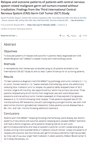
Relapse and outcome patterns of patients with central nervous system mixed malignant germ cell tumors treated without irradiation: Findings from the Third International Central Nervous System (CNS) Germ Cell Tumor (GCT) Study
Objectives
To evaluate patterns of relapse and outcome in patients newly diagnosed with CNS Mixed Malignant GCT (MMGCT) treated initially with chemotherapy alone.
Methods
A retrospective chart review was conducted using all 25 patients enrolled on the International CNS GCT Study III, with at least 7 years follow-up for all surviving patients.
Results
Thirteen patients at diagnosis had CNS MMGCT by pathology and tumor markers (n = 11), or tumor markers alone (n = 2). Twelve received chemotherapy alone, one additionally receiving focal irradiation prior to relapse. Six patients (46%) relapsed (mean of 30.5 months; range 6–59 months), two beyond and four within the primary site alone. Three patients relapsed early (6–23 months from diagnosis), two with alpha-fetoprotein elevations and one without tumor markers assessed; all three expired of progressive disease at 2–10 months following initial relapse. Three patients relapsed late (37–59 months) without AFP elevations, one with pathologically pure germinoma, two with mild beta-human chorionic gonadotropin elevations; these patients survive disease-free at 86+, 94+, and 126+ months following additional treatment.
Conclusions
Patients with CNS MMGCT relapsing following chemotherapy alone display two distinct patterns of recurrence and outcome; patients relapsing early possess MMGCT elements and have a dismal prognosis, while patients relapsing late do so with pure germinomatous elements and have an excellent outcome. Current cooperative group studies utilizing more localized fields of irradiation should monitor closely the patterns of relapse and outcome; late recurrences with germinomatous elements might be avoided by initial use of low-dose larger field irradiation in select patients. Pediatr Blood Cancer © 2015 Wiley Periodicals, Inc.
LINK: https://onlinelibrary.wiley.com/doi/abs/10.1002/pbc.25606
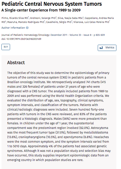
Pediatric Central Nervous System Tumors
The objective of this study was to determine the epidemiology of primary tumors of the central nervous system (CNS) in pediatric patients from a Brazilian oncology institute. We retrospectively analyzed 741 charts (415 males and 326 females) of patients under 21 years of age who were diagnosed with a CNS tumor. The analysis included patients from 1989 to 2009 and was performed using the World Health Organization criteria. We evaluated the distribution of age, sex, topography, clinical symptoms, symptom intervals, and classification of the tumors. Patients with clinical/radiologic diagnoses were included. Seven hundred forty-one patients with tumors in the CNS were reviewed, and 83% of the patients presented a histologic diagnosis. Males (56%) were more prevalent than females. In children under the age of 1 year, the supratentorial compartment was the predominant region involved (62.0%). Astrocytoma was the most frequent tumor type (37.0%), followed by medulloblastoma (13.6%), craniopharyngioma (10.5%), and ependymoma (6.8%). Headaches were the most common symptom, and the symptom intervals varied from 1 to 5010 days. Approximately 4% of the patients had associated genetic syndromes. Although it was not a population study and selection bias may have occurred, this study supplies important epidemiologic data from an emerging country in which population studies are rare.
LINK: https://journals.lww.com/jpho-online/Abstract/2011/12000/Pediatric_Central_Nervous_System_Tumors___A.7.aspx
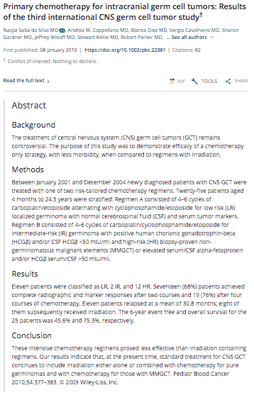
Primary chemotherapy for intracranial germ cell tumors: Results of the third international CNS germ cell tumor study
Background
The treatment of central nervous system (CNS) germ cell tumors (GCT) remains controversial. The purpose of this study was to demonstrate efficacy of a chemotherapy only strategy, with less morbidity, when compared to regimens with irradiation.
Methods
Between January 2001 and December 2004 newly diagnosed patients with CNS GCT were treated with one of two risk-tailored chemotherapy regimens. Twenty-five patients aged 4 months to 24.5 years were stratified: Regimen A consisted of 4–6 cycles of carboplatin/etoposide alternating with cyclophosphamide/etoposide for low risk (LR) localized germinoma with normal cerebrospinal fluid (CSF) and serum tumor markers. Regimen B consisted of 4–6 cycles of carboplatin/cyclophosphamide/etoposide for intermediate-risk (IR) germinoma with positive human chorionic gonadotrophin-beta (HCGβ) and/or CSF HCGβ <50 mIU/ml and high-risk (HR) biopsy-proven non-germinomatous malignant elements (MMGCT) or elevated serum/CSF alpha-fetoprotein and/or HCGβ serum/CSF >50 mIU/ml.
Results
Eleven patients were classified as LR, 2 IR, and 12 HR. Seventeen (68%) patients achieved complete radiographic and marker responses after two courses and 19 (76%) after four courses of chemotherapy. Eleven patients relapsed at a mean of 30.8 months; eight of them subsequently received irradiation. The 6-year event free and overall survival for the 25 patients was 45.6% and 75.3%, respectively.
Conclusion
These intensive chemotherapy regimens proved less effective than irradiation containing regimens. Our results indicate that, at the present time, standard treatment for CNS GCT continues to include irradiation either alone or combined with chemotherapy for pure germinomas and with chemotherapy for those with MMGCT. Pediatr Blood Cancer 2010;54:377–383. © 2009 Wiley-Liss, Inc.
LINK: https://www.nature.com/articles/ng.2645
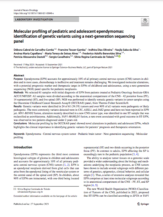
Purpose Ependymoma (EPN) accounts for approximately 10% of all primary central nervous system (CNS) tumors in chil- dren and in most cases, chemotherapy is ineffective and treatment remains challenging. We investigated molecular alterations, with a potential prognostic marker and therapeutic target in EPNs of childhood and adolescence, using a next-generation sequencing (NGS) panel specific for pediatric neoplasms.
Methods We selected 61 samples with initial diagnosis of EPN from patients treated at Pediatric Oncology Institute-GRA ACC/UNIFESP. All samples were divided according to the anatomical compartment of the CNS - 42 posterior fossa (PF), 14 supratentorial (ST), and five spinal (SP). NGS was performed to identify somatic genetic variants in tumor samples using the Oncomine Childhood Cancer Research Assay® (OCCRA®) panel, from Thermo Fisher Scientific®.
Results Genetic variants were identified in 24 of 61 (39.3%) tumors and over 90% of all variants were pathogenic or likely pathogenic. The most commonly variants detected were in CIC, ASXL1, and JAK2 genes and have not been reported in EPN yet. MN1-BEND2 fusion, alteration recently described in a new CNS tumor type, was identified in one ST sample that was reclassified as astroblastoma. Additionally, YAP1‐MAMLD1 fusion, a rare event associated with good outcome in ST-EPN, was observed in two patients diagnosed under 2 years old.
Conclusions Molecular profiling by the OCCRA® panel showed novel alterations in pediatric and adolescent EPNs, which highlights the clinical importance in identifying genetic variants for patients’ prognosis and therapeutic orientation.

Objetivo: Analisar as características epidemiológicas dos adolescentes portadores de neoplasias encaminhados para o Instituto de Oncologia Pediátrica (IOP/GRAACC) da Universidade Federal de São Paulo, entre os anos de 2000 a 2006.
Métodos: Trata-se de um estudo retrospectivo descritivo, em que foram avaliados os dados epidemiológicos dos pacientes, com idade entre dez e19 anos ao diagnóstico, admitidos do ano 2000 a 2006 no IOP/Graacc.
Resultados: Do total de 2.362 pacientes admitidos neste período com diagnóstico de câncer, 629 (26,6%) eram adolescentes. A idade média encontrada foi de 13,8 anos, sendo a maioria do sexo masculino (56,8%). Em relação à raça, 60,7% dos pacientes eram brancos. Os tipos de tumores mais frequentes foram: tumores de sistema nervoso central (22,1%), osteossarcoma (14,6%), linfomas (14,5%) e leucemias (14,5%). A sobrevida global, em cinco anos, dos 629 pacientes deste estudo foi de 73,7%. Destaca-se que os adolescentes com rabdomiossarcoma apresentavam doença disseminada e histologia de pior prognóstico, contribuindo para o aumento na taxa de mortalidade deste grupo de pacientes.
Conclusões: Os adolescentes com câncer correspondem a um grupo de pacientes que apresenta características peculiares quando comparado a outros grupos oncológicos.
Há diferença histológica dos tumores dos adolescentes com os da infância, em que predominam leucemias e tumores do sistema nervoso central. Nesse contexto, é fundamental facilitar o acesso desses pacientes a centros especializados e oferecer meios apropriados para o diagnóstico precoce e tratamento adequado.
Purpose: The purpose of this study was to assess, by panoramic radiographs, the prevalence of morphological dental changes in children with cancer who were submitted for chemotherapy alone or concomitant radiotherapy of the head and neck.
Methods: All patients admitted between March, 1996 and February, 2004 were analyzed and 137 were included in this retrospective, nonrandomized, institutional study. The rates of microdontia, taurodontia, anodontia, macrodontia, blunt root, and tapered root were assessed.
Results: The patients were distributed into 2 groups: (1) those with lymphoproliferative neoplasias (61%); and (2) those with solid tumors (39%). Their mean age when treatment began was 5 years and 6 months. Dental abnormalities were found in 39 (29%) patients, while 98 (72%) patients did not present any abnormality. The abnormalities found were: (1) microdontia (7%; N=10); (2) anodontia (6%; N=8); (3) taurodontia (14%; N=19); (4) macrodontia (5%; N=7); (5) blunted root (2%; N=2); and (6) tapered root (4%; N=5). Of these patients: 22% (N=30) presented 1 abnormality; 4% (N=6) presented 2 abnormalities; and 2% (N=3) presented 3 abnormalities.
Conclusion: Taurodontia was the most frequent abnormality found in children and adolescents who underwent antineoplastic treatment, and its rate was significantly higher than those found for the healthy Brazilian population. This study's results show that it is necessary for the odontologist to systematically research the dental changes that occur among this special group of patients.
LINK: https://www.ingentaconnect.com/content/aapd/jodc/2006/00000073/00000003/art00004

O objetivo deste artigo é relatar a experiência de implantação do modelo de prática avançada de enfermagem por meio da incorporação do enfermeiro clínico especialista na composição do quadro de enfermagem. As autoras apresentam o conceito desse modelo e descrevem seu processo de implantação em uma instituição de referência no cuidado à criança e ao adolescente com câncer da cidade de São Paulo, Brasil. Na avaliação das enfermeiras clínicas especialistas, o principal resultado obtido com a adoção do modelo refere-se ao fortalecimento do papel do enfermeiro como profissional de referência para a articulação das diferentes demandas de cuidado à criança, ao adolescente e ao adulto jovem com câncer e sua família. Também destacam a satisfação dos usuários com o estreitamento da relação paciente, família e enfermeira clínica, em todas as etapas do processo de cuidar.

Objetivo: Avaliar a frequencia de infeccao por Mycobacterium tuberculosis em criancas e adolescentes submetidos a TCTH ha mais de 1 ano, atraves do teste tuberculinico e do teste T-SPOT.TB, comparando-a a de um grupo controle sadio de idade semelhante; correlacionar a presenca da infeccao com caracteristicas do TCTH, nivel de reconstituicao imunologica pos-transplante e contato conhecido com paciente com tuberculose bacilifera. Desenvolvimento e metodos: Trata-se de um estudo transversal em que, de agosto de 2009 a junho de 2012, foram incluidos 114 individuos: 44 individuos saudaveis do grupo controle (C) (mediana de idade: 15,5 anos) e 70 pacientes submetidos a TCTH (T) (mediana de idade: 15,7 anos; mediana de tempo pos TCTH: 4 anos) acompanhados no IOP/GRAACC da UNIFESP. A avaliacao da infeccao por tuberculose foi realizada atraves de T-SPOT®.TB (Oxford Immunotech, Abdington, Reino Unido) e do teste tuberculinico, alem de radiografia de torax. A imunofenotipagem de linfocitos de sangue periferico foi feita atraves de citometria de fluxo. Para a analise estatistica utilizamos o teste Exato de Fisher e o Qui-Quadrado para as variaveis categoricas, e o teste Mann-Whitney para as variaveis quantitativas. Resultados: Dos 114 individuos, 2 pertencentes ao grupo transplantado e 1 individuo do grupo controle recusaram-se a fazer o TT, por isso foram excluidos da analise clinica e laboratorial para tuberculose. O grupo controle e o de pacientes transplantados foram comparaveis em relacao a idade, genero, classificacao socio-economica, presenca de marca da vacina BCG, contato intimo com tossidor cronico e frequencia de tuberculose latente (Grupo T= 5,8%, Grupo C= 9,3%; Exato de Fisher, p=0,708). A mediana do numero de celulas T CD3+/mm3 (Grupo T= 1468, Grupo C= 1722; Mann-Whitney, p=0,027), T CD4+/mm3 (Grupo T= 715, Grupo C= 1035; Mann-Whitney, p<0,001) e NK/mm3 (Grupo T= 209, Grupo C= 290; Mann-Whitney, p=0,002) foi menor no grupo dos transplantados. Apesar dessa diferenca, observou-se que a mediana de todas as populacoes de celulas analisadas entre os pacientes estava dentro dos valores de normalidade para a idade. Em cada grupo houve 4 casos de TT e/ou T-SPOT.TB positivos, com uma concordancia de mais de 90% entre os testes. A avaliacao destes 8 individuos com infeccao latente por M. tuberculosis mostrou que nao houve um padrao caracteristico de resposta a infeccao das celulas T CD4+, T CD8+ e T γδ em numero, e nenhum apresentou sintomas clinicos de doenca ativa. Conclusao: A frequencia de infeccao latente por Mycobacterium tuberculosis entre paciente pediatricos submetidos a TCTH foi comparavel a do grupo controle. Nenhum dos individuos do estudo apresentou tuberculose ativa. O teste tuberculinico mostrou-se um bom teste para o diagnostico da tuberculose latente, nao havendo a necessidade do uso de um teste confirmatorio in vitro nesses individuos que ja apresentam reconstituicao imunologica pos-TCTH.
LINK: https://repositorio.unifesp.br/handle/11600/22841

O objetivo do estudo foi avaliar a prevalência e a disseminação de amostras de Pseudomonas aeruginosa resistente aos carbapenêmicos e produtoras de metalo-β-lactamases isoladas de hemoculturas (2000-2005) de pacientes do Instituto de Oncologia Pediátrica da UNIFESP (IOP-GRAACC). MÉTODOS E RESULTADOS: Cinquenta e seis amostras de Pseudomonas aeruginosa foram isoladas de 49 pacientes. Trinta e duas dessas amostras foram classificadas como resistentes aos carbapenêmicos pela técnica de disco difusão e submetidas a reação de PCR para detecção de genes de MBL. Dezoitos dessas 32 amostras evidenciaram o gene blaSPM-1. Oito amostras selecionadas em diferentes anos no período de estudo apresentaram o mesmo perfil genético por pulsed-field gel electrophoresis. A terapêutica antimicrobiana foi considerada adequada em apenas 23,5% dos pacientes com bacteremia por P. aeruginosa carreando blaSPM-1 e letalidade de 70,6% no período de até 30 dias após a bacteremia e uma inadequação inicial dos esquemas antibióticos utilizados CONCLUSÕES: Evidenciamos a presença de um clone de P. aeruginosa resistente aos carbapenêmicos carreando blaSPM-1 que persistiu em amostras de hemocultura pelo período de 6 anos na instituição, com alta letalidade, justificando uma vigilância epidemiológica rigorosa e uma readequação dos esquemas de terapia antimicrobianos na instituição.
Resumo: Este estudo busca compreender a dimensão subjetiva de pacientes oncológicos que estudam ou
estudaram na escola hospitalar do IOP-GRAACC-UNIFESP. Através de uma metodologia qualitativa baseada
na Psicologia sociohistórica, há o objetivo de apreender os sentidos atribuídos pelos sujeitos estudados à
escola hospitalar, ao professor hospitalar e à aprendizagem que ocorre nesse contexto. Foi possível observar
que a escola hospitalar é marcada pela ausência significativa dos elementos que compõem o sentido acerca
da instituição escolar tradicional, ao mesmo tempo em que é vista como um espaço de aprendizagem
considerado mais agradável. O professor é carregado de uma carga afetiva bastante positiva, e o saber por
ele vinculado ganha aspectos diferenciados. A escola hospitalar deve se afirmar e pautar sua atuação nas
suas diferenças, mesmo quando baseadas na ausência, assumindo-se, assim, como espaço escolar específico.
O presente estudo tem como objetivo apresentar subsídios e idéias para a elaboração de propostas pedagógicas para outros contextos hospitalares, bem como contribuir para a formação de professores em ambientes diversos dos de salas de aula tradicionais. Foi utilizado como cenário de investigação o Projeto Escola Móvel/Aluno Específico no IOP/GRAACC/UNIFESP. Os eixos teóricos que fundamentam este estudo estão voltados para os aspectos do conhecimento e da formação de professores, mais diretamente a prático-reflexiva. Trabalhamos com diferentes abordagens qualitativas da qual participam os alunos/pacientes atendidos pelo Projeto, no período de março de 2000 até junho de 2002. Os procedimentos de coleta de dados foram a entrevista individual e a observação participante. As fontes de coleta de dados foram a entrevista, os relatos de aula, a escuta da comunidade, listas de pacientes diários fornecidas pelo hospital e a etnografia de textos. Os principais resultados sinalizam para a formação de professores em ambientes diversos dos de salas de aula tradicionais, para a necessidade de se investigar o meio de forma sistemática e organizada e então organizar as ações. Ressignifica-se dessa forma, com a prática alterada pela pesquisa, o conceito de cultura escolar. Sugere ainda fórum de discussão em pequenas comunidades de professores e pesquisa com classes-hospitalares que procurem ir além do resgate da auto-estima e possibilitem, ao longo do tempo, avaliar o desempenho dos alunos em suas escolas de origem. Aponta também as questões do professor consigo mesmo no que se refere a crenças pessoais, a preocupações de trabalho em equipes multidisciplinares, à aceitação de desafios impostos pelo meio, à crença na sua capacidade de ensinar e na de seu aluno aprender e à busca constante de formação.
LINK: https://leto.pucsp.br/handle/handle/10029#preview-link0
Hospital do GRAACC
Grupo de Apoio ao Adolescente e a Criança com C GRAACC
CNPJ: 67.185.694/0001-50
Rua Pedro de Toledo, 572
Vila Clementino – São Paulo – SP
CEP 04039-001
TEL + 55 11 5080-8400
SAC + 55 11 5080-8569
Desenvolvimento Institucional
Rua Sena Madureira, 415
Vila Clementino – São Paulo – SP
CEP 04021-051
TEL +55 11 5908-9100
Bazar
Rua Pedro de Toledo, 276
Vila Clementino – São Paulo – SP
CEP 04039-000